Uncommon Cause of Endotracheal Tube Obstruction during Anesthesia Recovery
- DOI
- 10.2991/dsahmj.k.191022.001How to use a DOI?
- Keywords
- Airway obstruction; anaesthesia; paediatrics surgery; recovery
- Abstract
Acute endotracheal tube (ETT) obstruction is not an uncommon event during surgery, but under certain circumstances, it can be life-threatening. It can arise for a variety of reasons. In this case study, we present an event of endotracheal tube obstruction in a pediatric surgery case during anesthesia recovery. Early detection and prompt response are the keys to successful management.
- Copyright
- © 2019 Dr. Sulaiman Al Habib Medical Group. Publishing services by Atlantis Press International B.V.
- Open Access
- This is an open access article distributed under the CC BY-NC 4.0 license (http://creativecommons.org/licenses/by-nc/4.0/).
1. INTRODUCTION
Intraoperative ventilatory failure is a critical event that has been associated with various causes. However, Endotracheal Tube (ETT) obstruction remains a difficult one to recognize. It can result from blood impaction, mucus plug, manufacturing defects, and tube malfunction [1]. Once the diagnosis of ETT obstruction has been reached, a prompt response is required to restore ventilation. Replacement of the ETT is a cornerstone in management. This case report describes a sudden ventilatory failure at the end of a surgical procedure in a child under general anesthesia. A blood clot obstructing the ETT was detected and successfully managed.
2. CASE REPORT
A 14-year-old girl (weight 45 kg, height 155 cm) was scheduled for cystectomy of left lung hydatid cyst under general anesthesia. There were no abnormal findings in the preoperative evaluation. There was no recent history of upper respiratory tract infection. Auscultation of the chest immediately prior to the surgical procedure revealed the presence of breath sounds bilaterally with no added sounds. Laboratory workup values (hematology and chemistry profile) were within normal range. Her Electrocardiogram (ECG) showed normal sinus rhythm. Apart from the left lung hydatid cyst, no other abnormal findings were identified on the chest X-ray scan.
The induction of anesthesia was smooth and uneventful, carried out with Intravenous (IV) propofol, fentanyl, and vecuronium. A Grade 1 laryngeal view was detected by direct laryngoscopy. A 6-mm cuffed ETT was used for intubation. Bilateral air entry on chest auscultation confirmed the ETT placement and followed by capnographic monitoring. The ETT was fixed at the 13 cm mark. The patient was then mechanically ventilated at a Peak Airway Pressure (PAP) of 15 cmH2O, and anesthesia was maintained with oxygen, nitrous oxide, and sevoflurane.
The surgical procedure was carried out in the right lateral position. Throughout the intervention, suctioning of the ETT was carried out several times to maintain airway patency. The surgical resection of the cyst proceeded smoothly with minimal blood loss. The patient was hemodynamically stable throughout the procedure. After closure of the wound, the patient was placed on the supine position, and a left side chest tube was placed to drain the pleural space. After the placement of the chest drain, drug administration was discontinued, and the patient had spontaneous ventilation with adequate chest rise and exhaled tidal volume of 200–250 ml. About 5 min later, despite being placed on 100% oxygen, the patient became distressed, exhaled tidal volume dropped to 60–100 ml, and SpO2 declined progressively to reach 79%. PAP reached 35 cmH2O and end-tidal CO2 (ETCO2) 70 mmHg. Upon auscultation of the chest, there was no wheezing, but air entry was diminished bilaterally. Fortunately, her blood pressure and heart rate were within the normal range.
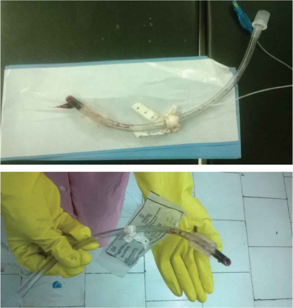
Introduction of the suction catheter was met with resistance and thus was unable to pass. When pulled out, the suction catheter was bloodstained. The patient was immediately ventilated with an ambo bag. However, resistance to manual ventilation was evident. At that time, we suspected acute obstruction of the ETT. Hence, the patient was extubated and oxygenated via face mask. Inspection of the tube revealed a blood clot occluding the air passage (Figure 1). Immediately after extubation, there were signs of improved compliance and chest rise. Hemodynamically, the patient was stable and the ETCO2 returned to normal. The patient woke up completely and there was no further incidence. She was transferred to the intensive care unit (ICU), where she spent the first night. The next day, she was transferred to the pediatric surgery ward. She stayed there and experienced no complications, and was discharged on postoperative Day 5.

Blood clot impacted at the end of the Endotracheal Tube (ETT).
3. DISCUSSION
Intraoperative ETT obstruction is not uncommon. However, it is important to differentiate it from other causes of intraoperative ventilatory failure such as bronchospasm, pneumothorax, and chest wall rigidity [2]. Hence, eliciting signs from physical examination and detecting abnormal patterns on capnographic monitoring are valuable tools to rule out other possible reasons. Our patient developed an increase in PAP and ETCO2. This was associated with a drop in the exhaled tidal volume and SpO2. Upon physical examination and monitoring of the respiratory and hemodynamic parameters, the possibility of bronchospasm or pneumothorax was deemed less likely. The resistance to passing the suction catheter raised the possibility of an ETT obstruction. The resistance felt in attempting to manually ventilate the patient reinforced our suspicion of an ETT obstruction.
The fact that the occlusion occurred after the procedure was carried out and when sedation was stopped, drives us to suggest that the active effort of expiration due to light sedation could have triggered blood impaction in the ETT. Another contributing factor is the type of surgery performed. Thoracic surgery has been reported to be complicated by blood clots occluding ETT. Thapa et al. [1] experienced an ETT obstruction by a blood clot in an 8-month-old infant who underwent emergency drainage and thoracoscopic decortication for a retropharyngeal and anterior mediastinal abscess.
ETT obstruction by a blood clot has been reported to be associated with patient’s repositioning. Lim et al. [3] described a case of ETT obstruction by a blood clot in a patient undergoing lumbar spine surgery after being put into prone position. Our patient experienced a similar scenario with the acute obstruction happening after repositioning.
4. CONCLUSION
Acute obstruction of the ETT is a life-threatening condition that needs to be recognized and managed swiftly. Anesthetists need to keep ETT obstruction at the back of their mind in cases of intraoperative ventilatory failure. In well-established settings, Fiberoptic Bronchoscopy (FOB) is the gold standard management strategy. It has the advantage of being a diagnostic and therapeutic tool. However, in the absence of FOB, anesthetists should have a high clinical sense and index of suspicion for cases of ETT obstruction.
CONFLICTS OF INTEREST
The authors declare they have no conflicts of interest.
AUTHORS’ CONTRIBUTION
SM conceived the idea, wrote the case, report and the reviewed manuscript. MH wrote the case report and drafted the manuscript.
ETHICAL STATEMENT
Written informed consent was obtained from the family of the patient. The patient’s name has not been mentioned, and there are no identifiers to link the patient to the report.
Footnotes
REFERENCES
Cite this article
TY  - JOUR
AU  - Sami Menasri
AU  - Mustafa Hussein
PY  - 2019
DA  - 2019/10/30
TI  - Uncommon Cause of Endotracheal Tube Obstruction during Anesthesia Recovery
JO  - Dr. Sulaiman Al Habib Medical Journal
SP  - 75
EP  - 76
VL  - 1
IS  - 3-4
SN  - 2590-3349
UR  - https://doi.org/10.2991/dsahmj.k.191022.001
DO  - 10.2991/dsahmj.k.191022.001
ID  - Menasri2019
ER  -
