
PO-10 A MULTI-MODALITY 4D SYSTEM FOR ANALYSIS OF THE AORTIC MORPHOLOGY AND FUNCTION FROM MR OR CT
- DOI
- 10.1016/j.artres.2014.09.016How to use a DOI?
- Open Access
- This is an open access article distributed under the CC BY-NC license.
Objective: Develop an integrated system for multi-modality 4D (3D+time) aortic data analysis including data construction, interactive pre-segmentation, accurate multi-surface segmentation and refinement, and quantitative index measurement. Provide a graphical user interface (GUI) with intuitive data visualization and workflow control. Test the capabilities of the system on MR and CT data.
Methods: 4D image with isotropic 3D voxels is constructed from DICOM images of a study. Candy-cane and LVOT MR data are fused together to benefit from their combined information. Lumen pre-segmentation is achieved by a few mouse clicks with instant feedback. Lumen (and outer wall for CT) surfaces are segmented in 3D by optimal graph search with embedded geometric constraints that guarantees to be global optimum. The result is refined using approximate clues instead of tediously redrawing individual contours. Quantitative indices – cross-sectional area, eccentricity and distensibility, centerline curvature and motion, wall thickness and calcification volume – are computed from complete 4D aortic surfaces. Due to its modularized design, each component can be independently fine-tuned for performance or extended to handle new modality or imaging protocol. The developed system provides simultaneous visualization of image, volume and surface data. The segmentation and quantitative analysis are guided by GUI with clear instructions and examples.
Results: Preliminary evaluation of segmentation accuracy on 20 dataset (10 MR, 10 CT) showed good absolute surface positioning errors (MR: 1.5±1.4mm, CT: 0.4±0.6mm). The user only needs to specify two aortic end points and several points inside the lumen on one 3D image. The computational time is approximately 20 seconds for 2.5GB 10-phase CT data or 500MB 20-phase MR data.
Conclusion: We develop a multi-modality 4D aorta analysis system with user-friendly interface and powerful segmentation and refinement tools. Various novel quantitative indices are computed from the complete 4D aortic surfaces, thus enabling comprehensive analysis of aortic physiology and morphology.
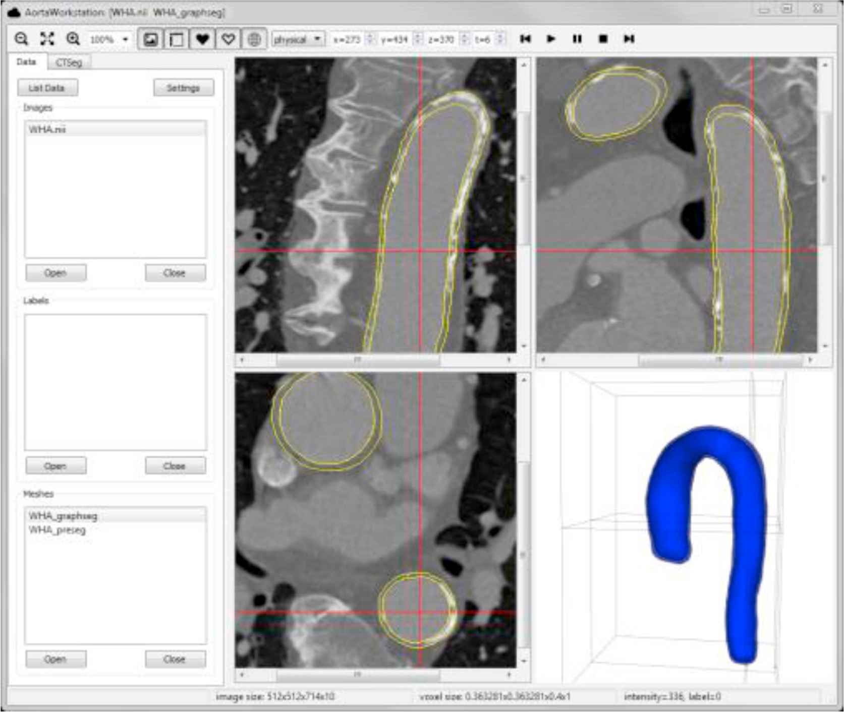
Example of data visualization: image data is shown on three orthogonal slices, segmented surface is shown by 3D rendering and its intersections with image slices (yellow contours).
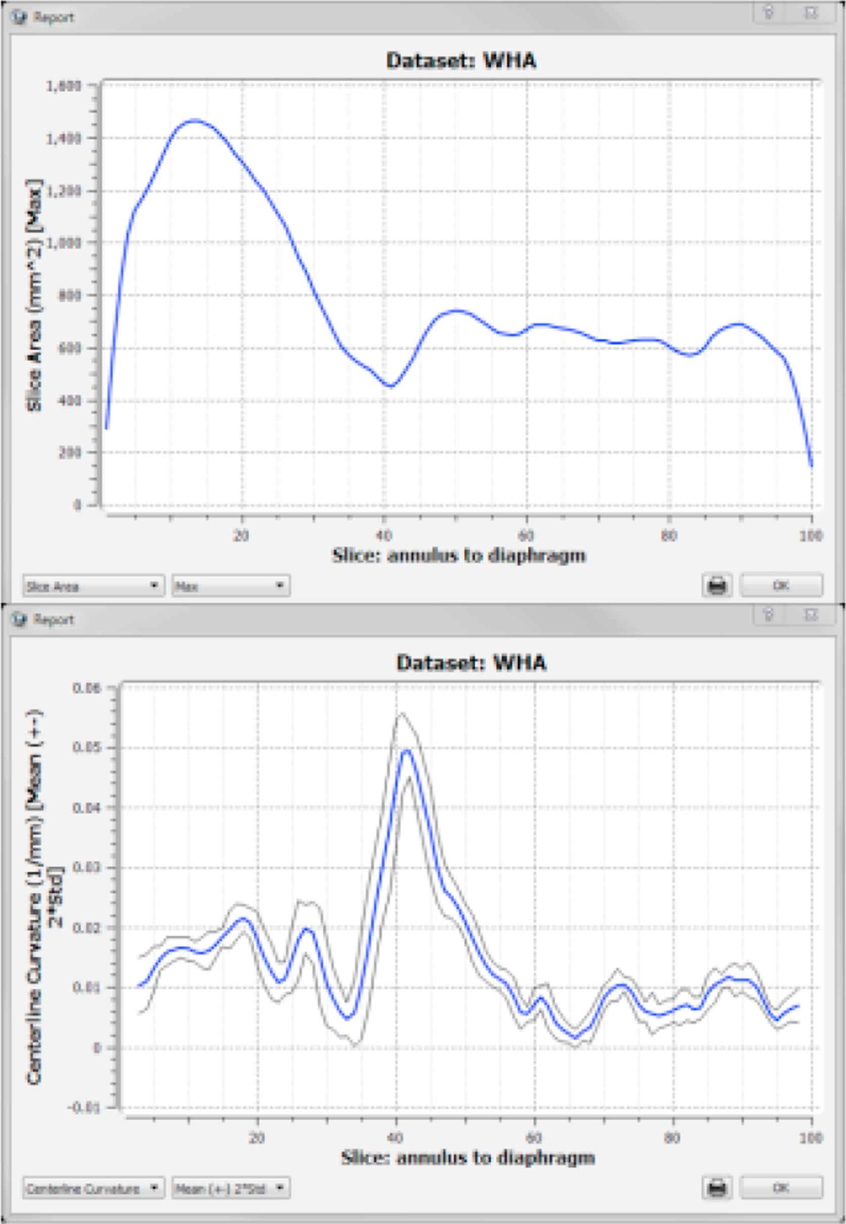
Examples of quantitative indices derived from a cardiac cycle. Top: maximal cross-sectional areas, bottom: range (mean and 2*standard deviation) of centerline curvature.
Cite this article
TY  - JOUR
AU  - Honghai Zhang
AU  - Xiangmin Zhang
AU  - Thomas Scholz
AU  - Gardar Sigurdsson
AU  - Andreas Wahle
PY  - 2014
DA  - 2014/11/04
TI  - PO-10 A MULTI-MODALITY 4D SYSTEM FOR ANALYSIS OF THE AORTIC MORPHOLOGY AND FUNCTION FROM MR OR CT
JO  - Artery Research
SP  - 169
EP  - 170
VL  - 8
IS  - 4
SN  - 1876-4401
UR  - https://doi.org/10.1016/j.artres.2014.09.016
DO  - 10.1016/j.artres.2014.09.016
ID  - Zhang2014
ER  -